
Photoacoustic Probes Enable Deep Brain Tissue Imaging
HEIDELBERG, Germany, Sept. 2, 2024 — A molecular engineering spearheaded by two groups at the European Molecular Biology Laboratory (EMBL) has developed an approach to create photoacoustic probes for neuroscience applications.
Scientists can learn more about biological processes by tracking certain chemicals, such as ions or biomolecules. Photoacoustic probes can act as ‘reporters’ for hard-to-detect chemicals by binding to them specifically. The probes can then absorb light when excited by lasers and emit sound waves that can be detected by specialized imaging equipment. For neuroscience applications, however, researchers have so far been unable to engineer targeted reporters that can visualize brain functions tailored for photoacoustics.
“Photoacoustics offer a way to capture imagery of an entire mouse brain, but we just lacked the right probes to visualize a neuron’s activity,” said Robert Prevedel, an EMBL group leader and a senior author on the paper.
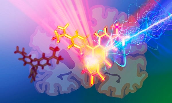
Newly developed photoacoustic probes allow scientists to explore deeper into the brain by enabling them to label and visualize neurons. Courtesy of EMBL/Isabel Romero Calvo.
To overcome this challenge, Prevedel enlisted the help of fellow EMBL group leader Claire Deo, also a senior author on the paper. She and her team specialize in chemical engineering.
“We have been able to show that we can actually label neurons in specific brain areas with probes bright enough to be detected by our customized photoacoustic microscope,” Prevedel said.
While researchers have experimented with using synthetic dyes as photoacoustic reporters of neuronal activity, controlling where the dye goes and what might be labelled has been challenging. Proteins have been particularly useful as probes for tagging specific molecules, but have not yet led to effective photoacoustic probes to monitor neural activity across the entire brain.
“In our case, we took the best of both of these sensors, combining a protein with a rationally designed synthetic dye, and we can now label and visualize neurons in specific regions of interest,” said Alexander Cook, first author of the study and a predoctoral fellow in the Deo group. In rational design approaches, researchers use existing knowledge and principles to build molecules with the desired properties, instead of blindly making and testing random compounds. The probe not only gave a static observation, Cook said, but showed a reversible, dynamic response to calcium, which is a marker of neuron activity.
According to Deo, an important challenge stood in the way of this technological development. Because photoacoustic probes have not been extensively studied, the researchers lacked a way to evaluate the probes they were building.
Consequently, the project began with Nikita Kaydanov, co-author of the study and predoctoral fellow in the Prevedel Group, who custom-made a spectroscopy setup.
“There is no commercial setup that can measure photoacoustic signals of a probe in test tubes or cuvettes, so we had to build one,” Kaydanov said. “We created our own photoacoustic spectrometer to assess and optimize the probes.”
“This allowed us to evaluate and characterize the different probes we made to assess a few things,” Deo said. “Did they produce a detectable photoacoustic signal? Are they sensitive enough? That’s how we inferred the next steps.”
Having proved that the probes could work in vial, the team took the work further by devising a way to deliver the probes into a mouse brain where they successfully detected photoacoustic signals from neurons inside the targeted brain regions.
“While we are excited about the progress, we need to be clear that this is just the first generation of these probes,” Deo said. “While they offer a very promising approach, we have a lot more work to do, but it’s a good first demonstration of what this system can enable and the potential it has in better understanding brain function.”
The researchers plan to continue development of the technology by improving the dye delivery system and confirming the ability to use them for dynamic imaging inside cells.
The research was published in the Journal of the American Chemical Society (www.doi.org/10.1021/jacs.4c07080).
Published: September 2024