Researchers from Aston University have developed a method to improve the accuracy of blood flow measurements in the feet of patients with Type 2 diabetes. The laser-based technique improves accuracy in detecting minute changes in microcirculation, and it improves on the method of laser Doppler flowmetry (LDF), which is commonly used to check circulation in the feet.
LDF gives measurements in the form of blood flow averages — which doesn’t always give the doctor enough information.
Changes at the microcirculatory level — involving the smallest blood vessels within the circulatory system — can affect whether tissue lives or dies. People living with Type 2 diabetes can be at risk of foot amputations due to circulatory complications caused by the condition.

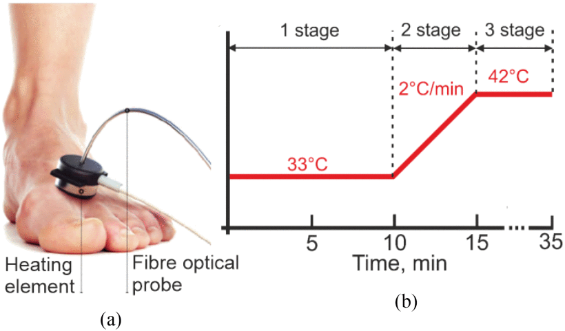
(a) Location of the fiber optical probe on the volunteer’s foot. (b) Study protocol with a heat test. Courtesy of Aston University, IEEE.
The researchers' approach changes the way that the light signals are processed, giving health care providers more information to work with. Currently, LDF systems look at blood perfusion, a quantity proportional to an average volume of blood flowing through an average volume of tissue per an average unit time.
The Aston method looks at rhythmic oscillations in microcirculatory blood flow called vasomotions; time series recordings of LDF signals allows these rhythms to be separated by origin and attributed to heart, respiratory, myogenic, neurogenic, and endothelial activity occurring within the patient. Underperformance of these rhythms can indicate developing complications of afflictions such as diabetes and rheumatic diseases.
By performing harmonic analysis of those rhythmic oscillations, the method is able to provide information with greater diagnostic significance.
With this technique, blood flow can be measured in a specific area of the vascular bed such as in the capillaries or veins.
The researchers validated the approach with standard occlusion tests conducted on healthy volunteers administered in the form of a probe on the surface of the skin. Limited clinical trials demonstrated that the approach can significantly improve the diagnostic accuracy of detection of microvascular changes in the skin of the feet in patients with Type 2 diabetes.
According to senior author Igor Meglinski, a professor of mechanical and biomedical engineering, the technique could be incorporated into most existing LDF systems.
In addition, the technique is not limited to feet. Meglinski noted a number of other potential applications, including functional brain imaging, diagnosis of vascular skin reactions triggered by allergens, and tumor angiogenesis.
Currently, the diagnostic capabilities, metrological certification, and scope of applications of the technology are still in the developmental stages. The next step, Meglinski said, which is already underway, is to collect and prove statistically the advantage of the technique.
The research was published in IEEE Transactions on Biomedical Engineering (www.doi.org/10.1109/TBME.2022.3181126).