Compiled by BioPhotonics staff
Sitting at a microscope for hours while
painstakingly searching for the right cell may soon become a methodology of the
past, thanks to software developed by researchers at the European Molecular Biology
Laboratory (EMBL).
The EMBL scientists have developed a computer program called Micropilot
that rapidly learns what the scientist is looking for and automatically performs
the complex microscopy experiment when it detects cells with particular features.
With the ability to incorporate machine learning, Micropilot can
analyze low-resolution microscopy images. Once it has identified a cell or structure
of interest to the scientist, it will automatically instruct the microscope to conduct
the experiment. The procedure can be as simple as recording high-resolution time-lapse
videos, or as complex as using a laser to interfere with fluorescently tagged proteins
and recording the results.
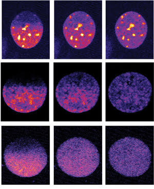
EMBL’s software Micropilot can detect cells at particular stages of cell division (each
row shows one cell). It instructs the microscope to remove fluorescent tags from
proteins in half the cell’s nucleus (left), and records what happens next
(middle and right). Courtesy of EMBL.
Eliminating the laborious and time-consuming task of searching
for cells, the software can easily and quickly generate enough data to obtain statistically
reliable results. In just four nights of unattended microscope operation, Micropilot
detected 232 cells in two specific stages of cell division and performed a complex
imaging experiment on them, a feat that normally would take a full-time experienced
microscopist at least a month. The observation should help scientists probe the
role of various proteins in a specialized biological process.
The EMBL teams that designed the software have used it to deploy
several types of microscopy experiments investigating different features of cell
division. They were able to detect when endoplasmic reticulum exit sites form, and
discovered the roles of two proteins, CBX1 and CENP-E, in condensing genetic material
into tightly wound chromosomes and in forming the spindle that helps align those
chromosomes. Their findings were reported online Jan. 23, 2011, in Nature Methods
(doi: 10.1038/nmeth.1558).