About This Webinar
This webinar will introduce the MUSE (Microscopy with Ultraviolet Surface Excitation) Microscope, a novel form of light microscopy that can generate high-quality histology and histopathology images directly from cut surfaces of fresh or fixed tissue samples of any thickness, with less than one minute of preparation. Presenter Richard Levenson, M.D., FCAP, will discuss the development of the MUSE microscope and demonstrate its use. He will provide a brief overview of light microscopy, including its history, use, value as a microscopy medium, and potential drawbacks.
Developed jointly by Lawrence Livermore National Laboratory and University of California, Davis (UC Davis), the MUSE microscope uses short-wavelength UV light which penetrates only microns-deep into tissue, eliminating the need for precision-cut, thin specimens. The UV light excites many fluorescent dyes simultaneously to provide detailed, high-resolution colored images.

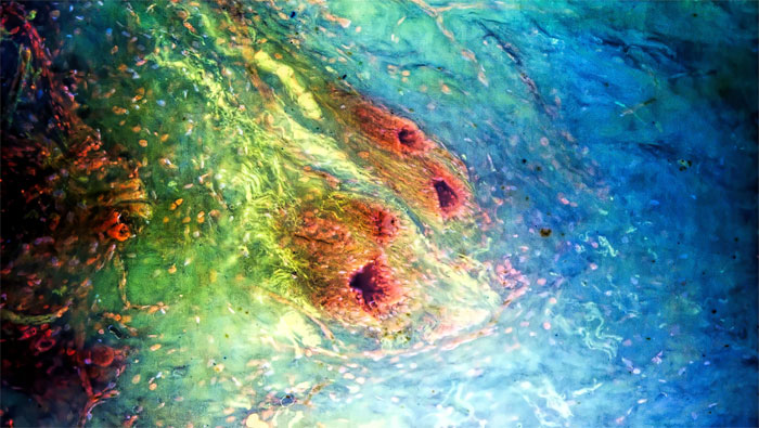
MUSE Microscope slide of breast ducts. Courtesy of Richard Levenson, M.D.
 About the presenter:
Richard Levenson is professor and vice chair for Strategic Technologies in the Department of Pathology and Laboratory Medicine at the University of California, Davis, and is co-founder and CEO of MUSE Microscopy Inc. He received his BA in History and Literature at Harvard College and MD at the University of Michigan. A pathology residency at Washington University, St. Louis, was followed by a cancer research fellowship at the University of Rochester, and an assistant professorship in pathology at Duke University. He became active in the optics field while at the National Science Foundation’s Center for Light Microscope Imaging and Biotechnology at Carnegie Mellon, and then joined Cambridge Research and Instrumentation, Inc. (now part of PerkinElmer), eventually as vice president for research. Following that, he consulted in optics, software, instrumentation and digital pathology before joining UC Davis in 2012.
Who should attend:
Researchers, scientists and clinicians who are interested in learning about novel forms of microscopy and recent advances in the field.
For more information on the MUSE microscope
read our story. Watch for an article on MUSE technology by Richard Levenson in the January issue of
Biophotonics.