About This Webinar
Alon Greenbaum discusses methods for quantitative analysis of intact organs using tissue clearing and adaptive light-sheet microscopy. As a test case, he presents macroscopic and microscopic properties of an intact porcine cochlea that is entombed by a dense and bony structure.
***This presentation premiered during the 2021
BioPhotonics Conference. For more information on Photonics Media conferences, visit
events.photonics.com.
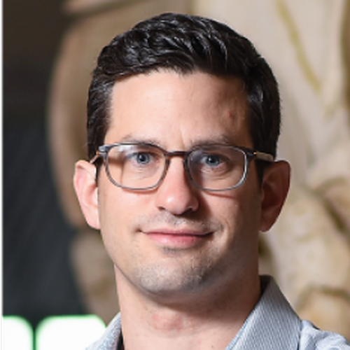
About the presenter:

Alon Greenbaum, Ph.D., joined North Carolina State University in January 2019 as a Chancellor's Faculty Excellence Program cluster hire in High-dimensional Integration of Biological Systems. An assistant professor in the joint University of North Carolina at Chapel Hill/NC State Department of Biomedical Engineering, Greenbaum develops complex imaging devices and algorithms to advance three-dimensional profiling of intact organs to answer biological questions regarding aging and disease progression. Application areas span the development of adaptive light-sheet microscopes and algorithms for rapid high-resolution imaging of whole organisms; computational tools to handle big data; and translational applications, such as exploration of rare stem-cell niches in the context of age-associated diseases. Greenbaum has been awarded the Good Ventures Postdoctoral Fellowship of the Life Sciences Research Foundation, as well as the Howard Hughes Medical Institute International Student Research Fellowship. He has published more than 25 peer-reviewed journal articles and his work has been presented at more than 30 conferences.