 Intraoperative OCT in Veterinary Surgery for Cancer
Tue, Aug 16, 2022 1:00 PM - 2:00 PM EDT
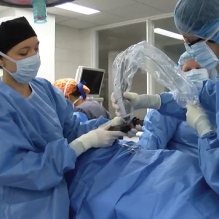
Surgery is a common cancer treatment performed in dogs and cats but the process of assessing the tumor takes several days and is only able to evaluate a small portion. Optical coherence tomography (OCT) is a non-invasive optical imaging technique that helps solve issues that accompany this process. OCT enables real-time intraoperative surgical margin assessment, allowing rapid visualization of the tissue microstructure at the surgical margins. To date, Dr. Laura Selmic, and her team have found high sensitivity and specificity for detection of incomplete margins after surgical excision of skin tumors, including STS and mast cell tumors, in dog and feline injection site sarcoma. The results reveal that OCT has potential for showing the demarcation between tumor and other normal tissues including muscle, fat, and skin.
|
|
|
|
|