|
Wednesday, November 2, 2016 |
|

|
|
|

|
November 2016
Spectroscopy Tech Pulse is a special edition newsletter from Photonics Media covering key developments in spectroscopy technology.
|
|
|
Near-Infrared Spectroscopy: Delving Deeper in the Brain
As a functional brain monitoring and imaging technique, functional near-infrared spectroscopy (NIRS) has undergone a tremendous transformation. Over the past two decades, the technology has grown from bulky prototype instruments measuring one or two points on the head to sophisticated systems providing whole-brain coverage and real-time display and enabling an array of important applications, including applications in behavioral research, psychiatry, memory loss, and stroke and brain injury. In many cases, the technological developments have occurred alongside changes in the user base and the emergence of new applications, often — though by no means always — in response to those changes.
|
|
|
|
|
|
Photosynthesis Map May Hold Clue to Improving Solar Cell Efficiency
The results of measuring the flow of energy during photosynthesis using spectroscopy could contribute to the development of more efficient solar energy technologies. Researchers at Lund University used ultrafast 2D electronic spectroscopy to track the excitation-energy flow through the entire photosynthetic system of green sulfur bacteria. They located the routes along which solar energy was transported, mapped the functional organization of individual complexes within the photosynthetic unit and demonstrated that energy was transferred within subunits within a period of subpicoseconds to a few picoseconds. They additionally demonstrated that across cell components, energy was transferred within a much lengthier timeframe of tens of picoseconds.
|
|
|
|
|
|
Two-Dimensional MEMS Arrays Pave Way for Mobile Spectrometers
In the field of near-infrared (NIR) spectroscopy, a system that combines portability with the accuracy and functionality of high-performance laboratory systems would significantly enhance real-time analysis. The development of small handheld spectrometers, powered by a battery, could lead to more efficient monitoring of industrial processes or assessments of food ripeness in the field.
|
|
|
|
|
|
Holographic Imaging Technique Diagnoses Malaria Automatically, Accurately
A quantitative phase spectroscopy (QPS) system that incorporates digital holography has been used to spot malaria-infected cells from a simple, untouched blood sample without any help from a human. The technique employs machine learning algorithms and has been shown accurate in detecting malaria infection 97 to 100 percent of the time. The research could form the basis of a fast, reliable test for malaria that could be given by most anyone, anywhere in the field.
|
|
|
|
|
|
Innovation in Surface Tracking Opens Doors to Raman Imaging Applications
Scientists dream of being able to probe variations in the chemical composition and structure of materials without any form of sample preparation. Raman spectroscopy, a noncontact, nondestructive analysis tool that yields information on the chemical, vibrational, crystal and electronic structure of materials at the submicron scale, promises this. The primary hurdle has been keeping samples in optical focus during imaging measurement.
|
|
|
|
|
|
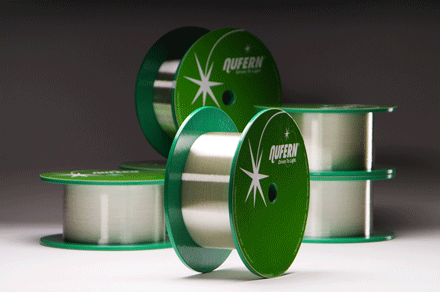 Nufern’s NuVIEW Optical Fibers
Nufern
The NuVIEW fiber family features specialty single mode and polarization maintaining fibers for the latest OCT, spectroscopy, and advanced medical imaging technologies. These fibers are designed to exceed the demanding requirements of todays advanced imaging systems. Request Info Visit Website
|
|
|
|
|
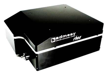 Rhea Ultra-Sensitive Spectrometer
Admesy BV
The Rhea series spectrometer offers a unique combination of ease of use and accurate measurement capabilities packed in a robust jacket. The Rhea utilizes a high-end cooled CCD detector for low noise and high dynamic range. Request Info Visit Website
|
|
|
|
|
|
Large-Scale, Deep-Tissue Neuronal Imaging
Wed, Jan 25, 2017 1:00 PM - 2:00 PM EST
Please join us for a free webinar! Lingjie Kong, Ph.D. will speak on advances in large-scale deep tissue imaging of biological dynamics, focusing on applications in neuroscience. Kong received his Ph.D. in Optical Engineering from Tsinghua University in 2012. For postdoctoral training, he worked at X. Sunney Xie's group at Harvard University and Meng Cui's group at Howard Hughes Medical Institute's Janelia Research Campus and Purdue University. He is currently engaged in research work at Purdue University and is planning to join the faculty at Tsinghua University.
|
|
|
|
|
 |
|