When you gaze into someone’s eyes, you can tell much about
their character. It just so happens that you also might be able to tell whether
they have a neurological disease.
Jacob W. Petrich and his colleagues at Iowa State University in
Ames have shown that basic noninvasive fluorescence spectroscopy can show whether
animals have been infected with a form of transmissible spongiform encephalopathy
(TSE). Experienced by cows as bovine spongiform encephalopathy (BSE, aka “mad
cow disease”) and by sheep and goats as scrapie, TSE is a neurodegenerative
disease that slowly destroys the brain via the growth and aggregation of protein-laden
structures called prions.
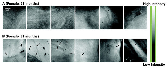
Hyperspectral fluorescence images can reveal whether an animal has a form of transmissible spongiform
encephalopathy. Shown are the retinas of sheep that are scrapie-free (A) and those
that have scrapie (B). Courtesy of the American Chemical Society.
An incurable malady related to Creutzfeldt-Jakob disease, BSE
can be transmitted from cattle to people, especially if infected cow brains and
spines are allowed to mix into the human food supply.
Using spectrofluorometry and hyperspectral fluorescence imaging
microscopy, Petrich’s team examined the eyeballs of several dozen sheep, looking
for evidence of scrapie in both infected and noninfected beasts. After observing
the spectra emitted from the various parts of each eyeball, the researchers found
that both the retina and the sclera (the protective tissue that covers most of the
eye) showed large differences in fluorescence intensity and other spectral features
between healthy and nonhealthy tissue.
The group’s members do not yet know the precise cause of
these differences, but they suspect that the prions cause an increase in a lipid-rich
material called lipofuscin, which allows the eyes to become a window to neurological
well-being.