Growing up, most people were probably told that dogs cannot see the world in quite the same way that the average human can —
perceiving the world in black and white
and shades of gray instead of the vibrant colors we enjoy.

Courtesy of iStock.com/HannamariaH.
But this provides only a partial picture. In actuality, canines’ eyes are a little more similar to human eyes than researchers originally conceived; they have similarities to ~8.5% of humans.
According to published research, dogs are color-blind but only red-green color-blind, meaning that they largely see shades of only yellow and blue, because they don’t have red cones in their eyes. This is not beneficial at stop lights, to be sure, but alright for other forms of visualization. For decades, scientists thought cone formation was a virtual coin toss and that the cells haphazardly committed to sensing green or red wavelengths. But a research team, led by Johns Hopkins University associate professor of biology Robert Johnston, found that the reasons may be more molecular.
Apparently, having wider visual access to the rainbow can be attributed to the presence of retinoic acid, a molecule derived from vitamin A. The presence of high levels of the molecule in the early development of organoids in vitro can be correlated with higher ratios of green cones found in the eye. By contrast, low levels of retinoic acid can be correlated with the generation of red cones. These ratios affect the protein opsin, which is found in all cone cells and is responsible for telling the brain which colors it can see. It does provide a new slant on the phrase “seeing red.”

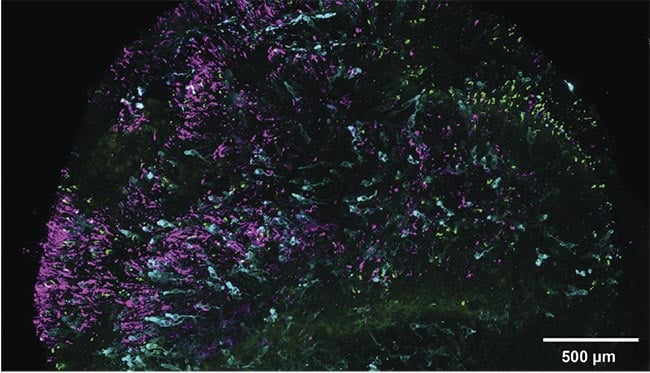
A retinal organoid used by a research team at Johns Hopkins University. Blue cones are marked in cyan and green and red cones are marked in green, while low-light perceiving rods are marked in magenta. Courtesy of
Sarah Hadyniak/Johns Hopkins University.
Though the amount of acid in each cone cell can cause each set of eyes to perceive wildly different things, the actual makeup of cell genes is 96% identical, which makes it hard to determine exactly when the divergence in development takes place. But, by growing a human retina in a petri dish and using colorimetric in situ hybridization to visualize opsin development, researchers were able to spot the genetic differences after tracking the cone ratio changes over the course of 200 days.
Testing also included the mapping of all cone cells in 700 adults to see whether there was a golden ratio between green and red cones. But because the ratio of green and red cones varied greatly between all the test subjects, the results were inconclusive.
While the researchers prepare to dive back into their investigation, literally through the eye of the needle, they are spurred on by their potential future findings with a goal to more fully grasp how cones and other cells link to the nervous system. They hope that future results lead to understanding diseases such as macular degeneration and glaucoma. Meanwhile, the rest of the world can fantasize about seeing the world through their pets’ eyes.