
Optical Redox Imaging Monitors Heart Cell Development
An optical imaging tool for monitoring the growth of human heart cells known as cardiomyocytes (CMs) could lead to a reproducible means to generate human induced pluripotent stem cell-derived cardiomyocytes (iPSC-CMs) for biomanufacturing. The imaging technique, along with various synthetic hydrogel substrates, was developed by a team at the University of Wisconsin-Madison.
CMs derived from iPSCs are a promising tool for combatting heart disease, which is a leading cause of death worldwide. IPSCs can be used to improve drug screening platforms, build accurate disease models, and develop personalized regenerative medical treatments. They can be reprogrammed to generate high-purity CM batches. However, in vitro maturation of iPSC-CMs remains difficult.
IPSC-CM differentiation and maturation studies typically rely on heterogeneous substrates and destructive verification methods. The technique that is traditionally used to grow iPSC-CMs is highly variable, prone to contamination, and degrades easily over time. The assays used to screen CM cells to determine their maturity often require harsh chemicals that damage the cells.
An optical imaging technique paired with hydrogel substrates provides an accessible, nondestructive approach to monitoring the growth of heart cells for biomanufacturing. Courtesy of the University of Wisconsin-Madison.
The University of Wisconsin-Madison team recognized that a touch-free monitoring approach and tunable substrates might provide a way to produce homogenous, functional CM cells and determine the ideal conditions for the cells’ successful growth.
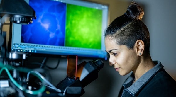
The researchers developed a label-free, wide-field optical imaging technique to observe live CM cells throughout their differentiation and early-to-late maturation and identify key metabolic shifts in the cells. They grew the cells on synthetic, in-house developed hydrogels.
Using autofluorescence lifetime imaging (FLIM), the researchers verified increased oxidative metabolism and NAD(P)H protein binding activity throughout cell differentiation. To increase throughput and screen more hydrogel formulations, they used widefield NAD(P)H and FAD autofluorescence imaging to calculate the optical redox ratio at multiple timepoints throughout differentiation. The optical redox ratio represents the metabolic shifts in the cells’ energy production and consumption.
The researchers developed several different compositions of synthetic, modifiable hydrogels to induce stem cell differentiation and maturation. Their approach to formulating the gels led to uniform results.
“The goal is having something that will be tunable and reproducible,” researcher Danielle Desa said. “The dream application of using these synthetic materials would be for biomanufacturing, because you’d want something robust and repeatable.”
The researchers found that the optical redox ratio decreased in CMs grown in all the synthetic hydrogel formulations during differentiation, early maturation, and late maturation. These results indicate that the CMs’ metabolic pathways shifted from glycolysis (i.e., breakdown of glucose to access energy quickly) to oxidative phosphorylation (i.e., use of oxygen to produce energy).
The team screened CMs grown in the hydrogels out to 100 days post-differentiation and continued to see metabolic changes over that period. “It was useful to see that they didn’t degrade in that time,” Desa said. “We wanted to do repeated imaging on the same gels over time and see if the technique was sensitive to any metabolic changes.”
The researchers also observed metabolic differences in low- and high-efficiency differentiation batches of cells. The sensitivity of the optical redux imaging technique could serve as an early predictor of which batch of cells is most likely to mature into functional CMs. This feature could make optical redux imaging a useful screening tool for cell manufacturing.
The efficient production and widespread use of iPSC-CMs will require consistent manufacturing of functionally mature cell batches and touch-free, nondestructive monitoring. The optical redux imaging technique and synthetic hydrogels could provide an accessible, medium-throughput method to monitor iPSC metabolism in response to interventions that aim to increase batch yield, consistency, and maturity without manipulating or destroying the cells. The technique allows the same cells to be monitored repeatedly and used for complementary analyses.
The research was published in Biophotonics Discovery (www.doi.org/10.1117/1.BIOS.1.1.015002).
Published: September 2024