NIR Spectroscopy Could Provide Window into AD Pathology, Therapeutics
BEDFORD, Mass., Sept. 1, 2021 — To detect Alzheimer’s disease (AD) in its early stages, researchers at the VA Bedford and VA Boston Healthcare systems developed a noninvasive optical technique that uses near-infrared spectroscopy to identify changes in the brain by capturing chemical and structural information from brain tissue.
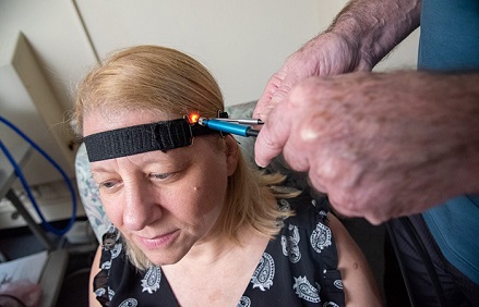
To acquire data, the researchers positioned two in-house-developed fiber optic probes at the patient’s temple, one next to the other. One probe delivered near-infrared light from the light source into the patient’s brain. The other probe collected the diffusely reflected light and delivered it to a detector. Using this approach, the researchers acquired diffuse scattering spectra from 650 to 1050 nm.
Because near-infrared light is weakly absorbed, it can go through several centimeters of tissue to reach the brain from the surface of the scalp. The light signal from the researchers’ spectrometer was able to interrogate a portion of the temporal lobe as well as overlaying tissues.
The researchers measured the effects of the light by comparing the light from the source optical fiber with the light collected by the detector fiber. The collected light differed from the light that entered the brain because of the way it interacted with the brain tissue.
In a previous collaboration with Boston University’s Alzheimer’s Disease Center, the researchers demonstrated their technology using tissue samples from autopsies. Using near-infrared spectroscopy on the autopsy samples, they differentiated between brains confirmed to have AD and those that did not.
In a new study, the researchers applied their technique to three groups of living volunteers: patients with mild cognitive impairment (MCI), age-matched healthy controls, and late-stage patients whose AD diagnosis was confirmed by an autopsy after they died. The three groups (MCI, CTL, AD) showed differences in the average intensity of diffusely reflected light.
A computer algorithm developed by the researchers enabled them to identify patterns in the spectroscopy data. By comparing the light refraction from healthy tissue with diseased tissue, they were able to identify specific refraction characteristics present in tissue affected by AD.
The researchers identified two spectral features in the regions of 860 and 895 nm that signaled a difference between patients. The slope variate at 895 nm played a more important role in distinguishing MCI subjects from controls, whereas the slope variate at 860 nm was more important for distinguishing MCI patients from those with AD.

Researchers at the VA Bedford Healthcare System demonstrate how near-infrared spectroscopy is used to detect brain changes possibly linked to Alzheimer's disease. Courtesy of Frank Curran.
Considering that the two slope variates were not correlated, the researchers suggest that the slope variate at 895 nm could be more significant earlier in the disease and that data at 860 nm could play a more important role later in the disease’s progression.
According to the researchers, the work marks the first successful classification of a neurodegenerative condition in vivo by near-infrared optical spectroscopy.
“This technology is significant because it probes the biochemical and cellular structures of the brain noninvasively with a technique that is inexpensive and could be put into widespread use,” researcher Eugene Hanlon said. “Most importantly, it gives useful information about those with mild cognitive impairment.”
In a best-case scenario, the signal at 895 nm would be found to respond to an intervention that prevented the progression of the 860-nm signal. In this best-case scenario, treatments to prevent AD could be developed and their effects documented before the onset of symptoms and before those symptoms became irreversible. If longitudinal clinical trial studies show that the near-infrared spectroscopy technique can be used to track disease progression, the researchers believe their approach could become a safe, noninvasive method for identifying AD, potentially at an early stage, and for assessing response to treatments in real time.
The new technology could be especially helpful for veterans.
“Veterans are more at risk for Alzheimer’s disease than the general population,” lead researcher Frank Greco said. “This technique has the potential to help identify what factors may increase that risk.”
The spectroscopy method has been accepted by the Food and Drug Administration as a protocol for possible clinical use, based on the results of clinical trials. To prepare for clinical trials, the researchers are refining the design of the probe and the specifications for the spectrometer, the software, and the interpretation of the output data.
The research was published in the Journal of Alzheimer’s Disease (www.doi.10.3233/JAD-201021).
Published: September 2021