Compact detectors, sources, and optics enable the use of imaging techniques such as endoscopy in applications ranging from ophthalmology to cardiology.
By Antonio Castelo
The miniaturization of components used in medical imaging has facilitated a range of groundbreaking diagnostics and therapeutics in modern health care. These imaging systems help to create detailed visual representations of the interior of tissues and organs for clinical analysis and medical intervention for a wide variety of diseases and chronic conditions. They can also play a critical role in guiding operations and evaluating treatment responses in proximity to the surgical suite.
A few microcamera modules designed for endoscopy. Courtesy of OptaSensor.
Photonics technologies have played a major role in revolutionizing this field. These technologies rely on light interaction with biological tissues to detect structural and functional information at the cellular and molecular levels. This capability is especially valuable in fields such as ophthalmology, cardiology, and neurology, where early detection and precise monitoring are critical for successful outcomes.
Each modern method of medical imaging based on a photonic technique can offer unique strengths in visualizing internal tissues. For its part, endoscopy enables real-time, color video imaging of internal body surfaces, commonly used for diagnosing and treating conditions in the gastrointestinal, respiratory, and urinary tracts. It provides direct visual feedback and allows for real-time procedural actions, although it is an invasive technique that may require sedation or anesthesia.
Other methods such as optical coherence tomography (OCT) provide high-resolution, cross-sectional images of tissue microstructures, allowing clinicians to detect early-stage abnormalities just below the surface of tissue. These capabilities are especially valuable in diagnostics in ophthalmology and cardiology. More recently, spectral imaging techniques have been prominent in areas including multispectral and hyperspectral cameras in dermatology, wound care, and surgery. These systems enhance traditional imaging by capturing data across multiple wavelengths of light, allowing for the identification of subtle composition or structural changes in tissues, and variations in the blood flow.
Small scope, big impact
The use of endoscopy for medical imaging dates to the early 19th century, when the French inventor
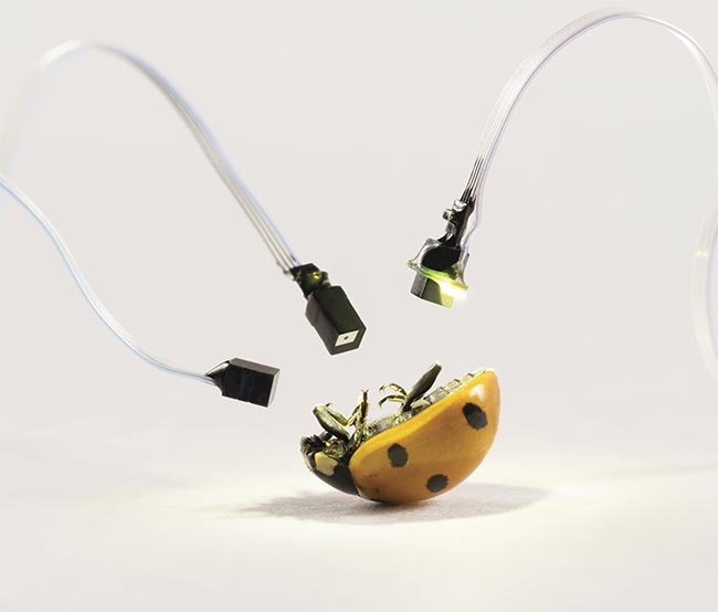
Antonin Jean Desormeaux used a device to guide chemical cauterization. The method experienced a huge improvement in the 20th century, with the invention of fiber optics and the integration of video cameras, permitting procedures to be viewed on monitors and recorded for subsequent analysis. For certain applications, endoscopes have evolved into thin, flexible tubes equipped with a light source and a camera (opening image). They can provide detailed, high-resolution images, allowing the early detection of diseases, such as ulcers or infections, without the need for major surgery. Its efficiency, accuracy, and lower risk compared with other exploratory procedures make it a valuable tool in modern medicine.
Miniaturization is a major trend in endoscopy, and is based on the development of smaller components for the emission, detection, and transmission of light. The goals are to obtain safer and more comfortable — not to mention more versatile — tools and access previously hard-to-reach areas in the body. A notable example of such miniaturized components is microcamera modules, which have become essential components in modern endoscopes. These modules enable high-resolution imaging within extremely small anatomical spaces.
The German company OptaSensor has been working in this field, manufacturing microcamera modules that combine compact image sensor chips with custom-designed lenses for small medical endoscopy applications (Figure 1). OptaSensor is introducing its OS500 camera, featuring a custom CMOS chip that incorporates advanced technology adapted from high-end imaging applications. It includes backside-illumination pixel technology and a through-silicon via chip-scale package. And, at the heart of the camera is a specially engineered wafer-level lens, developed by Opta-
Sensor and manufactured in Europe.
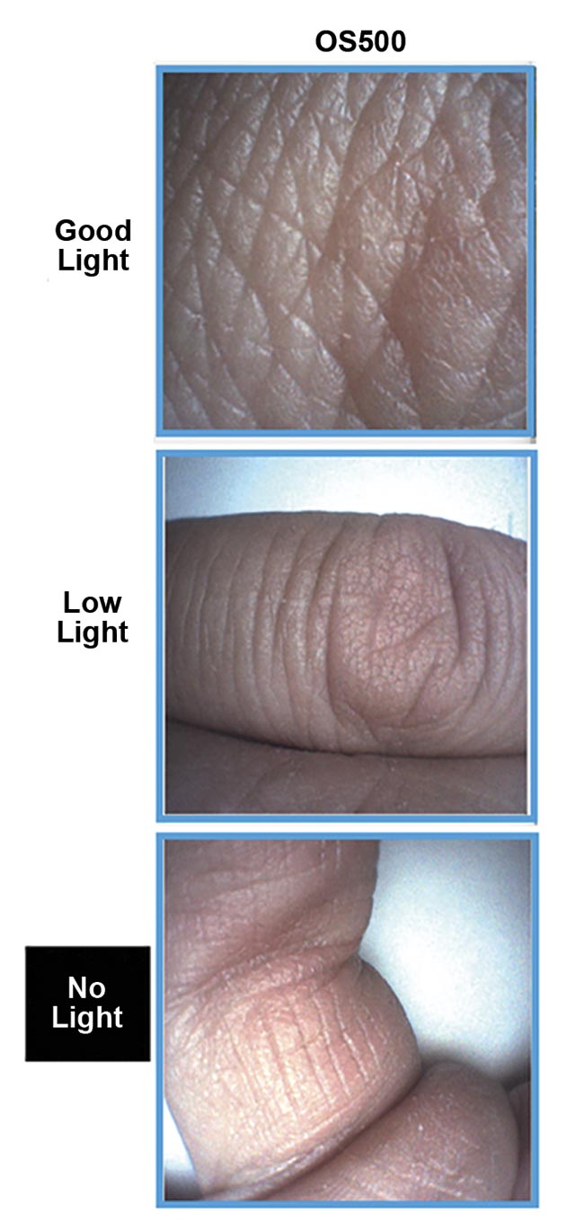
Figure 1. Examples of images obtained with the microcamera module OS500 for endoscopy applications. Courtesy of OptaSensor.
The OS500 camera was designed for use at the distal end of long endoscopes, because it can operate without requiring additional components. This helps to minimize module size and volume and has allowed the development of devices with tighter bending radii, an important benefit in certain medical procedures such as those in the vasculature or gastrointestinal tract. Moreover, the camera can be connected directly to microcontrollers using a serial peripheral interface-like communication protocol. This has potential for applications where the advantages of a microcamera are needed, but cable length is less critical, such as eye tracking. The OS500 can be customized with various sensor resolutions, lens designs, light filters, apertures, and integrated illumination rings.
Optical components must also be smaller and more compact for the development of endoscopes with reduced size. Mikrop AG is a Swiss company specializing in the production and assembly of miniaturized optical components and systems. It offers a comprehensive range of optical components tailored for endoscopic applications, including highly complex optical micro-objectives for multi- and single-use applications (Figure 2).
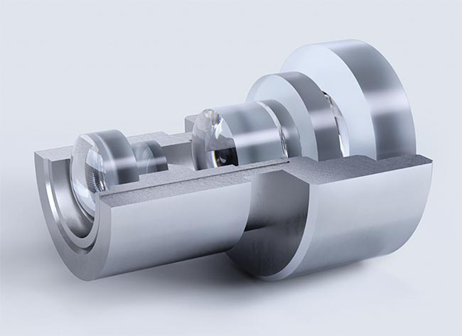
Figure 2. Micro-objectives for compact endoscopes. Courtesy of Mikrop.
These micro-objectives are designed to be integrated with image sensors, enabling high-resolution imaging in confined spaces. Mikrop offers glass micro-objectives with diameters of 1 mm with a field of view of up to 120° and a direction of view of up to 110°, with nearly diffraction-limited resolution. These components can safely withstand sterilization in an autoclave. For special and compact applications in high volumes, these plastic micro-objectives can provide similar optical performance. Microtechnology and the integration of components are also crucial in the process of the miniaturization of the endoscopes. For example, the German company Jenoptik develops highly sophisticated polymer and glass modules to integrate the optical, electronic, or mechanical components that are in these devices. Reliable and cost-effective volume production is possible as a result. In this process, European manufacturers such as OptaSensor, Mikrop, and Jenoptik have been supported by the International Microtechnology Business Network, which unites professionals in high-tech industries to bring innovative technologies and products to market.
OCT components
Certain photonics technologies have been adapted to provide increasing benefit in the medical arena, and the trend toward compact size and functionality has driven significant changes in these areas. OCT is a high-resolution imaging technique with established and expanding applications in medicine and biology. Some OCT systems, called time-domain OCT, use coherent near-infrared light to obtain micrometer-level depth-resolved images of biological tissue (particularly in the eye) or other scattering media. They measure the time delay of light echoes by mechanically moving a reference mirror.
More recent OCT methods emerged in the early 2000s, such as spectral-domain OCT, which uses a spectrometer to analyze interference patterns in the frequency domain, or swept-source OCT, where a swept laser source is needed to scan through different wavelengths. Advancements in sources, photonic components, and integrated optics have made it possible to design hand-held, wearable, and compact OCT devices.
Rapid improvements in tunable laser technology, especially external cavity lasers, allowed swept-source OCT systems to achieve high scan speeds and long imaging ranges. Additional recent innovations have been focused on improving spectral bandwidth, reducing noise levels, or expanding wavelength ranges to achieve higher resolution and deeper tissue penetration. For example, MEMS-
VCSEL technology offers a much longer imaging range, with a coherence length of >100 mm at A-scan rates of hundreds of kilohertz, but an imaging depth of 10 mm at megahertz A-scan rates. These characteristics enable full-eye imaging to improve cataract surgery and real-time imaging to aid in retinal diagnosis and surgery.
Advancements in sources, photonic components, and integrated optics have made it possible to design hand-held, wearable, and compact OCT devices.
The company OCTLIGHT ApS in Denmark, with their Eurostars project partners Wavesense Engineering and Medical University of Vienna, has been working on a dual-modality megahertz VCSEL-based swept-source OCT system for applications in ophthalmology. Defocus error — where an image is not in the correct position on a detection surface — is a major limitation of digital wavefront aberrometry. To address this, the Eurostars project partners have developed a modified system that allows for easy switching between fast anterior segment imaging and digital wavefront aberrometry with an extended defocus dynamic range of 20 D (Figure 3).
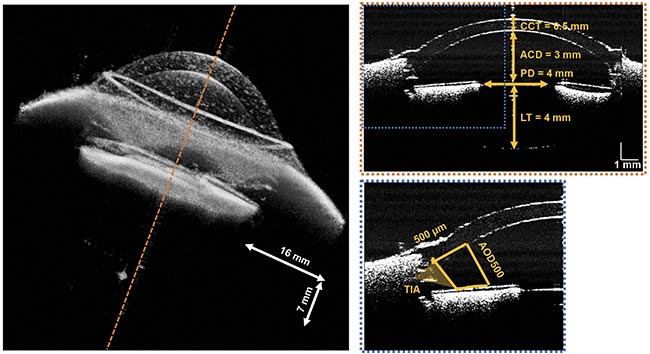
Figure 3. An OCT volume reconstructed from the average of upward and downward sweeps, showing a sample B-scan (selected at dotted orange line position) magnified at the dotted blue region of the anterior chamber of the model eye, recorded at a 1.6-MHz A-scan rate. ACD: anterior chamber depth; AOD500: the angle-opening distance at 500-µm anterior to the scleral spur; CCT: central cornea thickness; LT: lens thickness; PD: pupil diameter; TIA: trabecular-iris area. Courtesy of OCTLIGHT ApS and Wavesense Engineering.
Combining anterior segment imaging with aberrometry is beneficial in refractive error-related ophthalmic surgery and will support ophthalmologists in personalized planning of the surgical procedure. In comparison with other proposed solutions, the full speed of a 1.6-MHz swept laser centered at 1069 nm of the OCTLIGHT system facilitates faster and extended anterior imaging in depth, reducing the motion artifacts and improving wavefront sensing. With this solution supported by Eurostars, a fast anatomical imaging of the anterior segment of the eye has been demonstrated1.
Swept-source OCT systems can also benefit from the use of superluminescent diodes as light sources, because they offer a balance between the high spatial coherence of laser diodes and the broad spectral bandwidth of light-emitting diodes. This unique combination results in short coherence lengths, which are essential for achieving high-axial resolution in OCT imaging. The Irish company Superlum is active in this field, and works with academic and industrial institutions to develop effective gain chips for swept sources in wavelengths <1050 nm, down to the visible range of the spectrum.
In this region, swept-source OCT has advantages compared with other OCT techniques, particularly in terms of its imaging speed of large areas. In the context of the NETLAS (NExt generation of Tunable LASers) project, supported by the European Union, Superlum developed gain chips at 670 nm and 830 nm, which were successfully used for the demonstration of novel OCT swept sources at these bands. For medical imaging, the 670-nm swept source has been used by the University of Kent at Canterbury, and an extremely fast 828-kHz Fourier-domain mode-locking swept source at 840 nm has been used by the University of Lübeck.
The performance of spectral-domain OCT has also improved as a result of the development of new photonic components. For detection, they use spectrometers to measure interference patterns from reflected light without the need to mechanically scan the reference arm. The company Ibsen Photonics in Denmark is providing high-end spectrometers for this technique, with a strong focus in the ophthalmology market. Ibsen has developed advanced transmission grating technology, which offers high diffraction efficiency for OCT imaging wavelengths, paired with a diffraction-limited optical design of the spectrometers. Ibsen Photonics recently launched the EAGLE OCT series of spectrometers that covers the wavelength range between 800 and 890 nm with a resolution of 0.05 nm.
Such spectrometers are used in the diagnosis and monitoring of conditions such as age-related macular degeneration, glaucoma, and retinal detachment. Ibsen Photonics’ ongoing mission is to manufacture more compact and portable instruments and to expand to wavelengths that are used in other applications. Spectrometer layouts are becoming more compact through monolithic designs, where multiple components are integrated in close proximity to reduce the footprint; the use of CMOS detectors enables their compact form factor, lower power consumption, and improved noise performance. With these components, final spectral-domain OCT systems can be faster and smaller without sacrificing image quality.
The power of spectral imaging
Hyperspectral imaging has become increasingly relevant in medicine. This method offers a simple, noninvasive way to reveal biochemical signatures without the need for ionizing radiation, large machinery, or dyes to help differentiate between similarly colored tissues. Analyzing spectral data from human tissue provides key insights that can inform diagnostics and surgical planning.
The company Living Optics in England developed a portable visible-NIR video-rate spectral imaging camera that offers real-time snapshot hyperspectral images for a wide range of applications in medicine and health care. It is a 5-MP camera with a selectable field of view (6° to 29°), covering the wavelength range between 440 and 900 nm, with 96 bands for each spectral sample. Living Optics explored the use of this camera for shade selection in dentistry, as an alternative to the visual comparison using a shade guide. Accurate shade selection is a fundamental step in this field to match the shade of a restoration to the patient’s natural teeth and avoid subsequent costly rectifications. This hyperspectral camera was initially used to measure the reflectance spectrum of the typical shade guides and compute the CIE values to CIE LAB coordinates. These coordinates were compared with the ones obtained for an in vivo measurement of human teeth, so the closest match to the shade guides was obtained to help dentists in the restoration process (Figure 4).
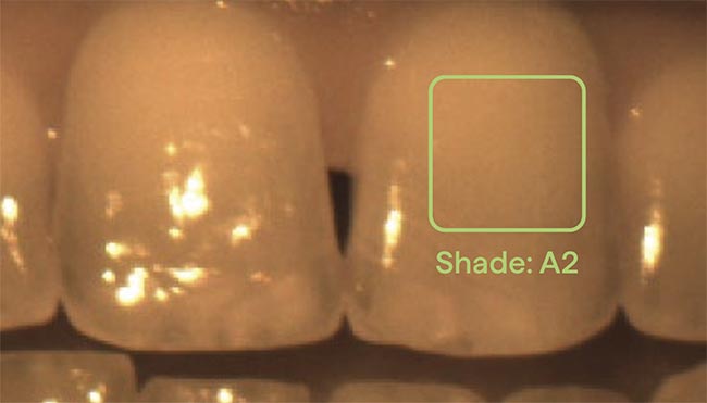
Figure 4. A hyperspectral image of a human tooth, annotated with the best VITA Classical shade match calculated for the rectangular region (green). Courtesy of Living Optics.
Multispectral imaging, a subset of hyperspectral imaging that captures a smaller number of bands, can also be highly valuable in medical applications. Several technological advancements in optics, sensors, and computing have led to the development of faster, more compact, and higher-resolution multispectral cameras. They have evolved into wearable, hand-held, or endoscopic systems, supporting applications in minimally invasive surgery, tissue oxygenation monitoring, and intraoperative guidance.
To improve the information obtained with the spectral images, Jenoptik developed its multispectral imaging MS-I2 solution, where illumination and imaging work in tandem for enhanced surgical outcomes. The complexity of modern surgical procedures demands a range of specialized illumination systems, each designed to meet the unique needs of specific treatments. The possibility of adjusting the light color on the fly ensures that every aspect of the surgical site is perfectly illuminated and allows the overlaying of critical images to detect even the smallest of details. By merging its competencies in illumination and imaging, Jenoptik developed a customizable OEM light engine that transforms multiple technologies into a single system and enables surgeons to perform complex procedures with more ease and accuracy.
All of these advancements in sensors, illumination, optical components, and on-board processing have led to significantly smaller and lighter systems that maintain high spectral resolution and imaging performance. They also broadly reflect that spectral imaging is moving toward mobility, integration, and cost-efficiency. Miniaturization is not only reducing size and weight, but also enabling wider adoption across emerging applications such as wearable health diagnostics and integration in surgical procedures.
Meet the author
Antonio Castelo is a physicist specializing in optics and currently serves as a technology manager at the European Photonics Industry Consortium (EPIC). After obtaining a Ph.D. in applied physics with a specialization in laser processing of materials, he worked as a sales engineer and sales manager for several value-added distributors and manufacturers. At EPIC, Castelo is in charge of the development of the laser and biomedical fields, and all related technology and applications. He has a strong interest in new technology involving photonics, such as ultrafast lasers, new medical devices, and spectroscopy solutions; email: antonio.castelo@epic-assoc.com.
Reference
1. M. Ke et al. (2024). Wide dynamic range digital aberration measurement and fast anterior-segment OCT imaging. Sensors, Vol. 24, No. 16, p. 5161.