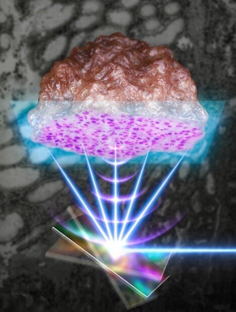
POHANG, South Korea, Nov. 4, 2021 — Researchers from Pohang University of Science and Technology (POSTECH) and Gachon University College of Medicine (South Korea) have developed a device that removes the need for complex procedures such as freezing, sectioning, and staining in histopathology process. The device is part of a system that uses ultraviolet microeletromechanical photoacoustic imaging microscopy (UV-MEMS PAM) technology.

Using a MEMS scanner that can scan the light and sound waves quickly, researchers in South Korea performed high-speed pathological biopsy on clinical tissue resected from cancer patients. Courtesy of POSTECH.
Cancer diagnosis is confirmed through histopathology by removing a part of the tissue in question, after tests such as MRI, CT, ultrasound, or endoscopy. Based on clinical diagnosis, the cancerous tissue is surgically removed and suspected tissues or lymph nodes are additionally examined. Medical personnel can than formulate treatment plans, and/or perform chemotherapy and radiation based on these results.
The machine learning-based histopathology approach uses a single-axis MEMS scanner as a novel label-free intraoperative histopathology method. In cancer resection surgery, histopathologic examination is essential to confirm the site of a tumor. Frozen-section testing has been conventionally used to carry out the examination, but due to its complicated process, it can prolong surgery and potentially cause interpretation errors.
Using a MEMS scanner in the newly developed approach, the researchers were able to visualize label-free cell nuclei of mouse and human tissues. By imaging clinical specimens resected from actual cancer patients and numerically quantifying the histopathologic results, the team demonstrated the potential of the proposed UV-PAM system as an alternative intraoperative histopathology method.
Photoacoustic imaging enables 3D imaging without an independent contrast agent. It additionally combines the advantages of high-resolution optical imaging with the ultrasound imaging that can achieve structural and functional imaging of small cells and living tissues all the way to large organs.
The microscope developed in the current work uses a high-speed MEMS scanner to significantly improve imaging speed, distinguishing normal tissues from cancer tissues by performing photoacoustic histopathology on cancer tissues that are excised from actual cancer patients, and quantifying the pathological microstructures, said professor Chulhong Kim, who leads the bio-optics and acoustics laboratory at POSTECH.
The research was published in Laser & Photonics Reviews (www.doi.org/10.1002/lpor.202100124).