Food Dye Curbs Light Scattering, Enables Live Tissue Imaging
The structure of biological tissues causes light to scatter, making optical imaging of the tissue difficult. Each biomaterial comprising the tissue, whether a fat, protein, or other type of biomolecule, has a different refractive index. The variety of refractive indices causes light to scatter as it passes through the tissue, making the tissue appear opaque. Also, the tissue absorbs light, which limits penetration depth.
A research team at Stanford University recognized that it was necessary to develop a method to match the different refractive indices within the tissue to allow light to travel through it unimpeded. The team applied fundamental optical principles to its hypothesis and developed a method that uses biocompatible, food-safe yellow dye to make living tissues transparent to visible light without increasing light absorption.

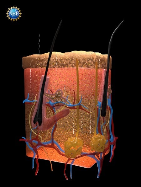
Illustration of skin tissues rendered transparent following saturation by Food, Drugs, and Cosmetics (FD&C) Yellow 5, including the paths of photons reflecting off undyed tissues. Courtesy of Keyi “Onyx” Li/U.S. National Science Foundation.
The new optical imaging approach could provide a straightforward, noninvasive way to help medical personnel diagnose a range of conditions, from injuries and internal disorders to cancer.
The researchers surmised that dyes that strongly absorb light could also be effective at directing light uniformly through different refractive indices. Using the Lorentz oscillator model for the dielectric properties of tissue components and absorbing molecules, they predicted that dye molecules with sharp absorption resonances in the near UV spectrum (300-400 nm) and blue region of the visible spectrum (400-500 nm) could raise the real part of the refractive index of an aqueous medium at longer wavelengths when dissolved in water. Thus, water-soluble dyes could effectively reduce the refractive index contrast between water and lipids, enabling optical transparency of live biological tissues.
The team found that tartrazine, an FDA-approved food dye commonly known as Food, Drugs, and Cosmetics (FD&C) Yellow 5, could match refractive indices and prevent light from scattering when it was dissolved into water and then absorbed into tissues. By tuning the refractive index of the surrounding medium to match that of the cells, the FD&C Yellow 5 dye made living tissues transparent.
The researchers conducted experiments in tissue-mimicking scattering hydrogels and ex vivo biological tissues. These tests confirmed the mechanism underlying the team’s observations and showed the achievable spatial resolution down to the micrometer level.
The team tested the dye on slices of chicken breast. As the tartrazine concentrations were increased, the refractive index of the fluid within the muscle cells rose until it matched the refractive index of the muscle proteins, and the tissue became transparent.
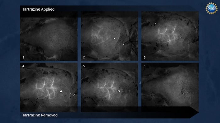
The researchers also tested a reversible tartrazine solution on live mice. They applied the solution to the abdomen, transforming the abdomen into a transparent medium that allowed for direct visualization of fluorescent protein-labeled neurons and captured the gut motility in the mice. The researchers also applied the dye solution to the scalp of a mouse head to visualize cerebral blood vessels and to the mouse hindlimb for high-resolution microscopic imaging of muscle sarcomeres.
When the dye was rinsed off, the tissues quickly returned to normal opacity. The tartrazine did not appear to have long-term effects, and any excess was excreted in waste within 48 hours. The researchers believe that injecting the dye could allow even deeper views within the tissues of an organism.

Time-lapse images of blood vessels in the brain just beneath the skull of a sedated mouse, revealed without any surgery, incisions, or damaging the mouse’s bone or skin. Courtesy of Stanford University/Gail Rupert/NSF.
The team applied two long-standing optical concepts to its research — Kramers-Kronig relations, which are mathematical formulas, and Lorentz oscillation, a phenomenon where electrons and atoms resonate within molecules as photons pass through. So far, these tools have not been applied to biomedical research in the way that the team used them. But the researchers found these concepts to be valuable for predicting how a given dye can raise the refractive index of biological fluids to match surrounding fats and proteins.
The researchers hope that their approach, which is grounded in fundamental physics, will launch a new field of study to match dyes to biological tissues based on optical properties.
“As an optics person, I’m amazed at how they got so much from exploiting the Kramers-Kronig relationship,” Adam Wax, program officer at the U.S. National Science Foundation, said. “Every optics student learns about them, but this team has used the equations to figure out how a strongly absorbing dye can make skin transparent.”
This approach to optical bioimaging offers a way to visualize the structure and activity of deep tissues and organs in vivo in a safe, temporary, noninvasive manner that could be used for a broad range of medical applications.
“Looking forward, this technology could make veins more visible for the drawing of blood, make laser-based tattoo removal more straightforward, or assist in the early detection and treatment of cancer,” professor Guosong Hong, a U.S. National Science Foundation CAREER grantee, said. “For example, certain therapies use lasers to eliminate cancerous and precancerous cells, but are limited to areas near the skin’s surface. This technique may be able to improve that light penetration.”
The research results also suggest that the search for high-performance optical clearing agents should focus on strongly absorbing molecules.
The research was published in Science (www.doi.org/10.1126/science.adr7935).
Published: September 2024