The unique properties of quantum dots allow them to be optimized for voltage sensing and for light-controlled electrical activation of cells.
Dr. Elena Molokanova, Joseph A. Bartel, Dr. Weiwen Zhao, Dr. Imad Naasani, Dr. Michael J. Ignatius,
Dr. Joseph A. Treadway, Dr. Alex Savtchenko, Invitrogen Corp.
Quantum dots are transforming life sciences imaging because of their extraordinary photostability, brightness, broad excitation, narrow emission, long fluorescence lifetimes and multiplexing capability. The nanometer-scale inorganic crystals contain a few hundred to a few thousand atoms of a semiconductor material, and their behavior is governed by the laws of quantum physics.
Quantum dots are not quite a molecule, although they have discrete electronic energy levels, and they are not a bulk semiconductor, either, although they demonstrate spin-orbit coupling and have large dielectric constants. Quantum effects become increasingly important as the physical size of a structure is reduced. The quantum confinement of light-generated excitons in three spatial dimensions determines the photophysical properties of quantum dots, such as the effective bandgap of the nanocrystals and, hence, the wavelength of fluorescence (Figure 1a).
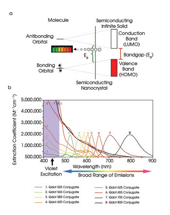
Figure 1. The size-dependent energy bandgap for semiconductor nanocrystals mirrors their emission (a). Quantum dots have a broadband absorption spectrum; thus, many colors of emission can be achieved using one excitation wavelength. The excitation (solid lines) and emission (dotted lines) spectra of quantum dot nanocrystals are shown (b). HOMO = the highest occupied molecular orbital; LUMO = the lowest unoccupied molecular orbital.
Quantum dots fluoresce differently than traditional fluorophores do. Absorption of any photons with energies higher than the bandgap causes the formation of excitons — or Coulomb-correlated electron-hole pairs. This broadband absorption spectrum means that quantum dots absorb light at every wavelength to the blue of their emission, and many colors can be obtained by exciting quantum dots of different sizes at a single wavelength to the blue of the bluest emission (Figure 1b). Electrons and holes stay separated for tens to hundreds of nanoseconds and then radiatively recombine, leading to the emission of a photon. The defined bandgap renders the emission spectra of quantum dots extremely narrow and symmetric.
Quantum dots are composed of hundreds to thousands of repeating atoms, each contributing to the nanocrystal’s ability to absorb light. Consequently, quantum dots have exceptionally high extinction (106 to 107 M–1cm–1), far greater than that of any organic dye. Moreover, optimally synthesized quantum dots possess quantum yields that approach the theoretical maximum of 100 percent. The combination of absorption properties with a large quantum yield makes quantum dots a thousand times brighter and more sensitive than organic fluorophores. These qualities often can reduce dramatically the concentration needed for biological applications and can heighten the ability to detect rare events. Additionally, the high photostability of quantum dots enables long-term imaging experiments under conditions that would cause the photoinduced deterioration of other types of fluorophores.
Quantum dots are highly engineered materials containing several structurally distinct elements. Although a nanoparticle’s core determines its color, a nanoparticle shell determines the brightness, photostability and environmental insensitivity. Proper deposition of an inorganic shell to the quantum dot cores produces a composite core-shell structure with enhanced chemical and photophysical properties.
The most common quantum dots are composed of CdSe and have emission wavelength maxima from ~400 to ~650 nm, which vary as the size of the nanocrystals is varied from 2 to 7 nm. A ligand added to the surface of the bare core-shell nanocrystal allows it to be dispersible in water and reactive toward biofunctional modifications, thus making it useful for biological applications. The resulting nanoparticles are 15 to 20 nm in size, which is similar to the size of GFP molecules. The fully assembled quantum dots are insensitive to the external environment and, therefore, are inherently more quantitative as reagents for macromolecule detection.
Because of their unique properties, quantum dot nanocrystals provide a platform for multiplexed fluorescence-based detection of unparalleled power and simplicity. These nanocrystals are used for bright, photostable and multicolor cell and tissue imaging, flow cytometry, Western blotting, single-molecule detection, in vivo imaging and more.
Monitoring functional activity
Although visualization of biologically relevant molecules in living cells is important, a better understanding of cellular homeostasis in health and disease requires new fluorescent probes that can track activities in intracellular signaling pathways with spatial and temporal fidelity.
Each cell has a resting membrane potential that originates from the separation of charges across its phospholipid bilayer. Changes in membrane potential are transmitted into cellular responses for various types of proteins, including voltage-gated ion channels, voltage-dependent phosphatases and G protein-coupled receptors. These proteins control a number of key processes, including neuronal signaling, heartbeat, brain function, muscle contraction, synaptic transmission, cell proliferation and hormone secretion.
Sensing and reporting changes in membrane potential is crucial for understanding transmembrane signaling. Electrophysiological methods, because of their high information content, are the gold standard for monitoring cellular electrical activity; however, optical methods have several advantages. For example, they are noninvasive and allow recording from multiple cells at a time as well as from tissue slices or intact organs.
Quantum dots are great candidates for voltage-sensitive probes because their physical size is comparable to the thickness of the cell membrane and because their electronic properties make them potentially tunable to the external electromagnetic field. A membrane potential of 100 mV across the hydrophobic lipid bilayer creates an intense local electric field reaching 107 V/m. This large field could interact with free charge carriers (electrons and holes) generated in quantum dots during illumination, thus changing their optoelectronic properties (Figure 2a).
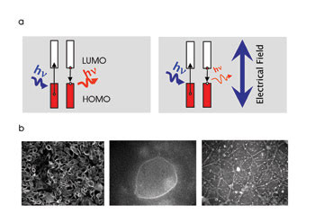
Figure 2. A change in membrane potential across the lipid bilayer of a cell creates an intense local electric field that can interact with the free charge carriers generated by excited quantum dots, making the latter usable as voltage sensors (a). Specific membrane labeling of CHO cells, NG108 cells and a neuronal network (left to right) using quantum dot-based voltage sensors is shown (b).
Because of their intentionally maximized environmental insensitivity, commercially available quantum dots would not suffice for this voltage sensing. This application would require the structure of nanoparticles to be engineered in a way that allows changes in the electric field to be transduced into changes in quantum dot emission. This sensitivity could be achieved by changing the absorption efficiency or the exciton behavior or by modulating an emitted photon. In addition, to sense membrane potential changes with maximum efficiency, quantum dots must be in or very near the cell membrane, within the highest transmembrane electrical field gradient. Ensuring intramembrane positioning requires a specialized, novel coating (Figure 2b).
We are developing quantum dot-based optical voltage sensors to monitor rapid changes in cell membrane potential. When inside the cellular membrane, the nanoparticles detect changes in transmembrane voltage gradient and report it as a change in their emission properties. During this project, we designed more than 100 combinations of inorganic structures and coatings based on our understanding of the physicochemical properties of semiconductor nanoparticles. Design variations have extended from various core compositions and shell structures to next-generation shapes. To find the most suitable nanomaterials for voltage-sensing applications, we performed screening experiments that addressed the specificity of membrane labeling and the sensitivity of nanostructures to changes in cell membrane potential.
In our screening experiments, cells in 96-well plates were incubated with quantum dot-based voltage sensors for 1 h, and then any excess nanoparticles were washed away. A population of labeled cells was depolarized with a chemical stimulus (100 mM KCl) — appropriate for high-content imaging experiments. Several selected quantum dot-based voltage sensors have exhibited ~0.5 percent change per 1 mV in fluorescence intensity in response to a depolarizing stimulus (Figure 3A). Standard electrophysiological protocols allow direct control of the cell membrane potential via a voltage-step depolarization delivered through a patch pipette to a cell in whole-cell mode. During these experiments, we observed that the fluorescence response of quantum dot-based voltage sensors correlated with electrophysiological recordings of the membrane potential (Figure 3b). Thus, quantum dot-based voltage sensors exhibit the sensitivity within the range of the physiological membrane potential and bring to cellular imaging added advantages such as photostability, large Stokes shift and multiplexing capability afforded by the unique optical properties of semiconductor nanoparticles.
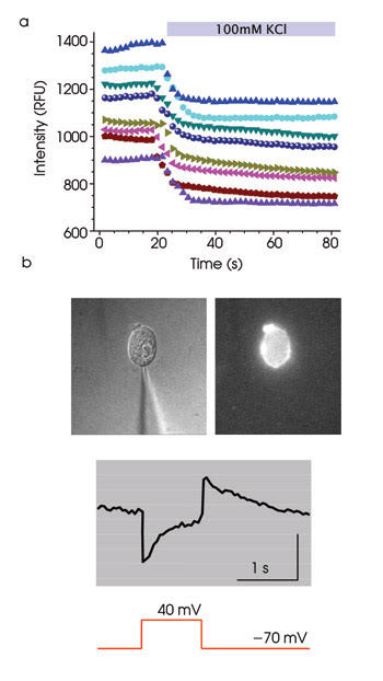
Figure 3. Quantum dot-based voltage sensors optically respond to chemical and electrical stimulation of cells. In multiple CHO cells, the fluorescence intensity of quantum dot-based voltage sensors changes in response to “high K+”-triggered membrane depolarization (a). A glass pipette was used for electrical stimulation of cells labeled with quantum dot-based voltage sensors (b, top). The fluorescence intensity of quantum dot-based voltage sensors’ response to electrophysiological stimulation is shown (b, bottom).
Besides passive monitoring of the cell functional activity, noninvasive manipulation of the membrane potential of cells is vital for understanding their development, their communication methods and their fate. Besides the direct stimulation of cells with microelectrodes, the most common approach is pharmacological intervention via the addition of “high K+” solution or pharmacological channel openers. These nonphysiological methods have serious shortcomings, including the lack of control of the membrane potential, irreversibility of elicited changes and low temporal resolution.
Light is a perfect trigger for remote reversible manipulation of the membrane potential in a temporally precise and spatially resolved manner. For example, genetically encoded light-sensitive proteins, such as naturally occurring channelrhodopsin (ChR2) or chemically modified ion channels, increasingly are being adopted in both in vitro and in vivo applications, despite concerns regarding the need for a high level of expression of exogenous proteins in the cells of interest.
Quantum dots may present an alternative solution for light-controlled electrical activation of live cells. They can be applied in a method similar to that used in photovoltaic applications, where quantum dot-based structures convert light to electricity. Upon exposure to the light, quantum dots can generate free electrons and holes. These free charge carriers either radiatively recombine and emit light, or they escape and create an electrical current.
Just as quantum dot nanoparticles are engineered to maximize radiative recombination (fluorescence), a next generation of particles will maximize current. When such quantum dots are formed into a three-dimensional array, strong electronic coupling between them produces excitons with a longer lifetime and facilitates the collection and transport of photogenerated free charge carriers. The rates of photogenerated carrier separation, transport and interfacial transfer across the contacts to the biological interface all must be fast to maximize the output. When trying to improve the photoconversion efficiency, it is important to keep in mind the efficiency of charge separation and the facilitation of the charge transport through the nanostructured platform.
To address the need for a light-controlled activation platform for biological applications, we exploited the photoelectronic properties of quantum dots and developed a nanostructured biocompatible interface composed of stacks of semiconductor nanoparticles coated with an adhesion layer. When these nanoparticles are placed in close proximity to a cell and are illuminated by visible light, the cumulative electromagnetic field generated by photoexcited nanoparticles (or light-induced current) modulates the cell membrane potential (Figure 4). This platform allows repeated physiological stimulation of cells and is compatible with any fluorescent readout because semiconductor nanoparticles can be excited by any light shorter than their emission wavelength.
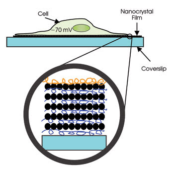
Figure 4. Quantum dots can be used for light-controlled electrical activation of live cells, as seen in this schematic, if a quantum dot-based activation coverslip is prepared.
To prepare multilayered quantum dot thin films for cell activation, we experimented extensively with quantum dots assembled on glass coverslips using a layer-by-layer deposition method. We also optimized the adhesion layer composition to achieve the closest contact between cells and activation substrate and, thus, increase the efficiency of conversion of light energy to changes in cell membrane potential.
In our experiments, cells were cultured on poly-L-lysine-coated nanoparticle-based glass coverslips for three to 14 days. Over the course of these experiments, we did not observe any adverse effects of nanoparticles on cellular morphology, on differentiation or on physiological responses. Depolarization of cells was achieved by passing the “activation” light from a mercury lamp through the bottom of a coverslip. In our experiments on a primary culture of hippocampal neurons, we used a current-clamp configuration to record the changes in membrane potential elicited by light. Light exposure of neurons cultured on activation coverslips resulted in a membrane depolarization that triggered the action potential (Figure 5a).
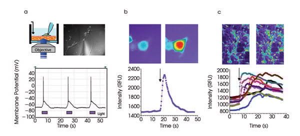
Figure 5. The experimental scheme and bright-field image of hippocampal neurons cultured on a quantum dot-based activation coverslip is shown (a, top). Changes in membrane potential and action potential are repeatedly triggered by light (a, bottom). NG108 cells loaded with fluo-4, a fluorescent calcium indicator, are shown before and after the activation light pulse (b, top). Changes in intracellular calcium concentration result from the light-triggered action potential (b, bottom). Hippocampal neurons with fluo-4 are shown before and after the activation light pulse (c, top). Changes in intracellular calcium concentration can be seen in several neurons as a result of light-triggered activation of neuronal network (c, bottom). The activation light pulse (380 nm) is identified with an arrow.
In summary, we demonstrated that, upon light illumination, our activation platform can repeatedly depolarize the membrane in nonexcitable cells (CHO cells, RBL cells) and can generate action potentials in excitable cells, such as NG108 cells and primary culture of hippocampal neurons with the only limitation being the gigaseal stability.
Because these nanoparticle-coated activation coverslips are transparent, they are compatible with various optical interrogation methods. We have designed an integrated optical assay that combines optical stimulation (via activation coverslips) and optical recording of cellular activity (via fluo-4 calcium indicator). We have performed these experiments both on NG108 cells and on primary culture of hippocampal neurons and have demonstrated the increase of intracellular calcium concentration as a result of light-triggered activation of cells. Figure 5b represents the calcium influx in NG108 cells through voltage-gated calcium channels involved in the light-triggered action potential.
Figure 5c demonstrates the heterogeneous activation of the neuronal network by light. Here some neurons were activated directly by light (note the fast-rising calcium flux), whereas other neurons were activated indirectly through interneuronal connections (note delayed and, sometimes, multipeak calcium flux pattern). Thus, this nanocrystal-based activation substrate promises to deliver a physiologically relevant activation stimulus compatible with optical methods of registration. Potential applications include functional studies of voltage-sensitive membrane proteins; studies of communications between cells in dissociated cell culture or tissue slices, including synaptic plasticity; cardiology studies; activation-stimulated cellular expansion and differentiation of stem cells.
In summary, using quantum dots as imaging probes to detect signaling cellular pathway components is just the first step in realizing the particles’ full potential. Novel biological functions for quantum dots, such as voltage sensing and changing cell membrane potential, advance them beyond the “lightbulb” applications — a direction in life sciences imaging that we believe will continue.
Meet the authors
Elena Molokanova is a senior scientist, Joseph A. Bartel is a scientist, Weiwen Zhao is a senior scientist, Imad Naasani is a senior scientist, Michael J. Ignatius is a director, Joseph A. Treadway is a technical area manager, and Alex Savtchenko is a principal scientist at Invitrogen Corp. in Eugene, Ore.; e-mail: [email protected].