Scanners, filters, deformable mirrors, and other adaptive optics integrated into the microscope improve performance for neuroscience and intravital research studies.
LARS SANDSTRÖM, G&H GROUP
Confocal and multiphoton microscopy are commonly used to provide 3D-resolved images of in vitro or in vivo tissue samples without physical sectioning. But there are also inherent trade-offs in the use of these techniques when it comes to imaging depth, resolution, and speed. Recent developments in the use of acousto-optic and other photonic components integrated into a microscope are beginning to address these limitations in capability, opening up new opportunities in life science research.
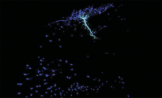
A place cell (cyan) and its local circuit of interneurons (dark blue) in a mouse hippocampus. Courtesy of Tristan Geiller/Losonczy Lab/Columbia’s Zuckerman Institute.
In confocal microscopy, a continuous-wave laser beam is focused into a small (micron-wide) beam waist inside a tissue sample containing a fluorescent dye or fluorescent protein. The resultant fluorescence passes back through the microscope objective and is then focused on a pinhole aperture located in front of a high-gain photodetector. This confocal aperture blocks any light that does not originate from the xyz location of the laser beam waist. By scanning the beam waist and/or moving the sample, a horizontal or vertical image slice or even an entire image cube can be acquired, with fluorescence captured at multiple depths.
Multiphoton microscopy is a related technique in which an ultrafast laser is focused using high-numerical-aperture optics. The laser wavelength is set to twice the wavelength needed for conventional excitation of the target fluorophore. At the beam waist, and only at the beam waist, the focused peak intensity exceeds the threshold for two-photon excitation. This provides inherent 3D resolution and eliminates the need for the lossy confocal aperture. Today, there are many variants of multiphoton microscopy based on other nonlinear optical effects that only occur at the beam waist.
Limits of conventional methods
Confocal and multiphoton applications are often negatively affected by practical trade-offs in imaging, such as in the capability to image deeper within tissue at the frame rates necessary to capture metabolic processes. In simple terms, speed can be limited by how fast the laser beam waist can be scanned relative to the sample (and vice versa). Traditionally, limitations have also existed in the modulation of the laser intensity, such as in the modulation needed for blanking during raster scanning.
Meanwhile, the resolution can be negatively affected by optical aberrations due to the microscope optics or, more insidiously, by the optical properties of the sample tissue itself, which may also be inhomogeneous. Fortunately, the innovative use of active photonic components is helping to address these limitations.
Acousto-optic deflectors
In confocal microscopy, the light beam is usually raster scanned in the xy plane, often using a pair of mirrors (one for each axis, x and y), each mounted on a galvanometer scanning coil. The fastest way to implement this approach is called resonant scanning, by which the galvanometers are driven to resonate (oscillate) back and forth at high speed. However, this has certain limitations, including nonlinearity of the motion, which is sinusoidal to a first approximation. Ideally, the beam should be scanned at a constant velocity for uniform pixel intensity. Recently, confocal and multiphoton microscope builders and users have attempted to avoid these speed and nonlinearity limitations by scanning the laser using acousto-optic deflectors (AODs) instead of galvanometers.
In an acousto-optic configuration, an acoustic wave is applied to some type of optically transmissive glass or crystal through which the laser light passes. For visible light applications, the acoustic wave is in the radio-frequency regime. This causes photoelastic deformations of the material, resulting in a periodic modulation of the optical properties, including the refractive index.
An AOD is configured to use this effect as a transmissive diffraction grating. As schematically illustrated in Figure 1, >90% of the incident laser beam undergoes first-order Bragg diffraction, where it is deflected from its original direction at an output angle determined by the radio frequency and other fixed factors. Thus, sweeping the radio frequency results in sweeping the output angle. This provides a much faster scanning method than galvos do, since the response time is limited only by the few nanoseconds or less that it takes the radio frequency to transit the active crystal aperture. This high speed is advantageous in the capture of cellular dynamics or neural activity.
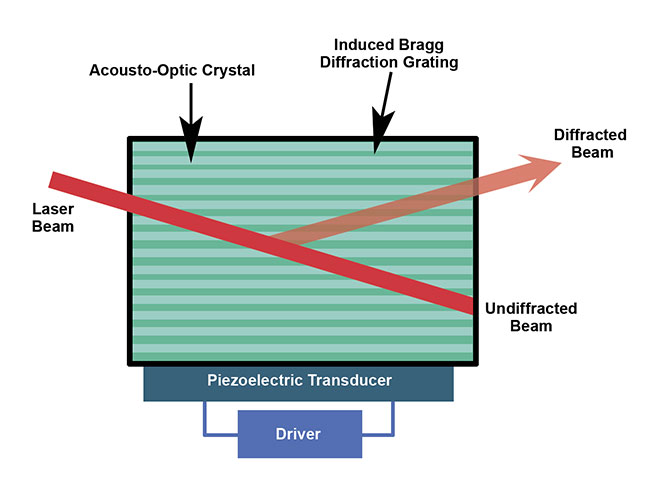
Figure 1. In an acousto-optic deflector, a piezoelectric transducer creates a Bragg grating in the
material. An aperture allows only the diffracted beam to pass. Courtesy of G&H.
A recently published study outlines research in which a multiphoton microscope was equipped with AODs for xy scanning. The study revealed important new insights in neuroscience, thanks to the combination of high speed and high resolution provided by AOD scanning. Specifically, the research group of professor Attila Losonczy at Columbia University has been the first to create a 3D inventory of interneuron activity deep in the mouse hippocampus1. This phenomenon was observed by imaging the calcium activity in the hippocampus in 3D in real time, with a high spatiotemporal resolution, using the AOD scanning multiphoton microscope.
The microscope used was a FEMTO3D Atlas from Femtonics, which actually uses four AODs — two for the x axis and two for the y axis — and it can perform various 3D region-of-interest scanning patterns for fast neuronal activity measurements and photostimulation. This research upended dogma by showing how so-called place cells — or neurons that code memory information about location — unexpectedly communicate with a network of excitatory neurons (See first image).
Beam control
The acousto-optic effect has been shown to also be beneficial through the creation of a quite different type of photonic component called an acousto-optic tunable filter (AOTF). This device acts as a dynamic (user-tunable) bandpass filter.
An AOTF uses birefringent material, which is a crystalline material in which the refractive index depends on the polarization of the incoming light beam. A radio-frequency actuator bonded to the side of the crystal causes acoustic waves that modulate the refractive index of the crystal material as in an AOD. Again, this AOD is configured to diffract most (>90%) of the incident light into a first-order deflected beam, which leaves the device through an aperture. But by using a birefringent material and a different internal geometry, the AOTF selectively deflects only one wavelength because of a phenomenon called phase matching.
The end result is a tunable device for which changing the radio frequency changes the wavelength at which the phase-matching condition is met. And if the crystal is cut and aligned correctly, changing the radio frequency does not change the angle at which the diffracted (and now wavelength-filtered) light leaves the device. The AOTF is thus a transmissive optic that acts as a wavelength-tunable narrow bandpass filter (Figure 2).
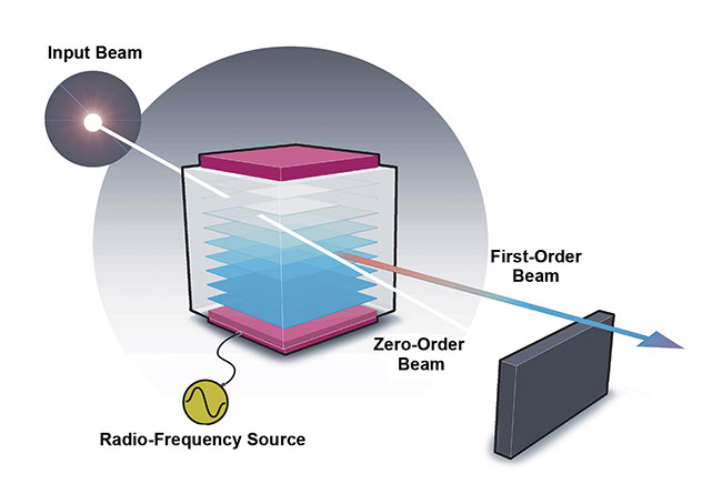
Figure 2. An acousto-optic tunable filter acts as a tunable bandpass filter in which the peak transmission wavelength depends on the frequency of the applied radio-frequency power. Courtesy of G&H.
Now, it may seem incongruous to use a wavelength tunable device in a laser-based technique. But the AOTF can actually be used in three different ways that can increase the speed of confocal microscopy. Many microscopes today are configured with multiple laser wavelengths from a multiline laser or a multilaser engine. First, the AOTF can be used to perform fast switching between excitation wavelengths, and it can also be used on the imaging side for fast switching of emission wavelengths. This allows for the analysis of multiple components in a sample.
Experiments often involve samples with more than one fluorophore, in which the emission from the sample is directed into different cameras using dichroic, cut-off, and bandpass filters, or into a single camera with a filter wheel. In the second example, the AOTF allows the use of a single camera with user-controlled dynamic wavelength tuning, again with adjustment taking only nanoseconds.
The third use of the AOTF in a confocal microscope system is for fast control of the laser power. Specifically, by changing the power of the radio frequency, the user can control the overall diffractive power of the device and hence the amount of the incident laser light that is diffracted into the first order and the exit aperture of the AOTF. (The AOD is often used for fast power modulation too, again by changing the radio-frequency power.)
The performance of acousto-optic devices, including AODs and AOTFs, is very dependent on the quality of the optical crystal element as well as on the quality the radio-frequency power source. These factors affect the maximum transmission of all acousto-optic devices and the transmitted wavefront quality, as well as factors such as the out-of-band extinction of an AOTF. These parameters, in turn, affect the microscope performance in terms of speed and image signal-to-noise ratio, particularly when two or more devices are used in the same microscope. For this reason, manufacturers place an emphasis on vertical integration — by growing their own crystals in-house.
Reflective adaptive optics
Neuroscientists are increasingly looking to image throughout the murine cortex, which has a thickness of ~1 mm, and even into the neocortex, which controls sensory perception and cognitive capabilities. Unfortunately, both the speed and resolution of confocal microscopy suffer due to the limitations of traditional optomechanics for scanning the z-axis (image depth), particularly for deeper images.
Dynamic scanning or stepping of this axis is usually implemented by physically moving the sample or microscope objective, which limits speed. This need to move the sample or objective can be a major problem if the microscope user wants to take images of tilted or uneven planes in the sample. And as the microscope is used to image deeper into tissue, aberrations caused by the tissue properties and the peculiarities of the microscope itself cause reduced resolution and blurring of the images.
One approach to correcting aberrations in deeper images borrows a proven concept from astronomy and uses deformable mirrors as adaptive optics. In astronomy, aberrations due to the atmosphere can compromise the resolution of ground-based telescopes. The latest telescopes use adaptive mirrors. With this configuration, mechanical actuators locally bend the primary mirror to optimize an image of a laser guide star, which is a point source created in the upper atmosphere by laser excitation of naturally occurring traces of sodium ions. Microscope users increasingly use microscopic fluorescent beads embedded in tissue to provide the same type of feedback in 3D imaging. And in microscopy, just as in astronomy, deformable mirrors can provide a dynamic solution for performing the aberration correction.
As shown schematically in Figure 3, small commercial mirrors specifically packaged for this type of application are based on a continuous reflective surface that is deformed by magnetic actuators. Importantly, they feature large strokes. This means these adaptive optics can not only correct for large spherical aberrations, they can also shift the depth of focus, providing random access, while maintaining a constant distance between the objective and the observed object. Figure 4 shows a set of well-resolved images of deep tissue acquired using these adaptive optics.

Figure 3. Large-stroke adaptive mirrors use tiny magnetic actuators to provide dynamic control of the mirror figure. Courtesy of ALPAO.
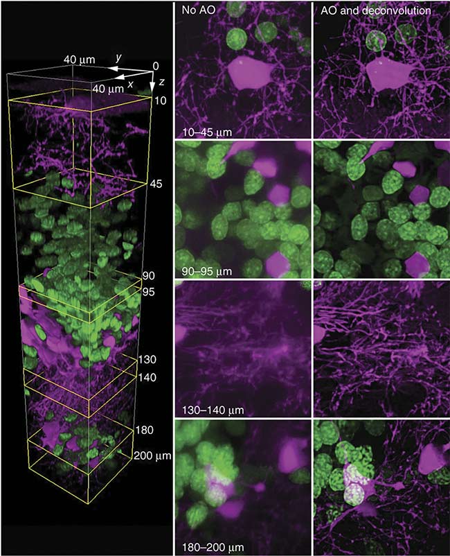
Figure 4. Adaptive mirrors and a descanned two-photon guide star provide a two-color confocal image of a living zebrafish brain (left). Image quality deteriorates with increased depth (middle). But even at 200 µm, the use of adaptive optics helps maintain high resolution (right). Courtesy of Kai Wang/Betzig Lab/Janelia Farm/HHMI.
Transmissive adaptive optics
Although adaptive mirrors offer extremely high reflectivity, and hence high efficiency, one drawback of using these mirrors in a microscope is that the beam path has to be specifically modified (folded) to accommodate the optics. An alternative adaptive-optic approach to dynamic wavefront correction involves a transmissive device, called a deformable phase plate, that avoids this complexity. This unique type of waveform modulator was recently developed by Phaseform GmbH in Freiburg, Germany.
The deformable phase plate acts as a type of adaptive liquid lens whose shape details (or “figure”) can be modified in real time under computer control. As shown in Figure 5, the deformable phase plate consists of a compact, sealed liquid-filled volume with a flexible polymeric membrane on one side and a rigid, transparent glass substrate on the other. An array of individually addressable transparent electrodes on the glass substrate cover the clear pupil area. The flexible membrane is supported by a micromachined spacer. The volume between the membrane and the substrate is filled with a high-refractive-index liquid. The conductive membrane is pulled toward the substrate when a voltage signal is applied to the electrodes. This actuation displaces the liquid and changes the effective optical path length of light that refracts through it.
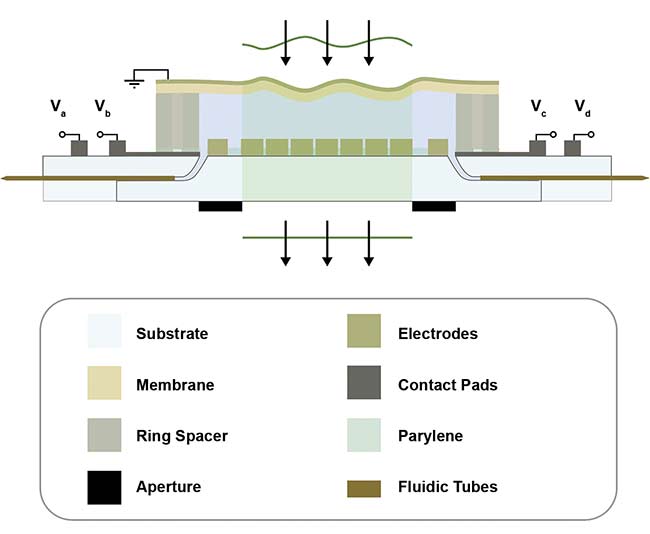
Figure 5. A schematic cross-section view of a deformable phase plate shows how it works as an adaptive lens. Courtesy of Phaseform GmbH.
Due to the slim design of the deformable phase plate, the device produces almost no dispersion, and multiple devices can be stacked on top of each other. This has applications in everything from microscopy in the life sciences to AR/VR.
Addressing speed and depth
Acousto-optics can provide yet another fast-focusing solution, thanks to a device called an acousto-optic lens. The acousto-optic lens concept has been around for over 20 years, but early formats had significant drawbacks, particularly for continuous-wave laser excitation. And while the use of early acousto-optic lenses enabled fast z-scanning and random xyz sampling using pulsed lasers (such as in multiphoton microscopes), the acousto-optic lenses create their own spherical aberration issues, limiting the resolution for deeper tissue studies.
Recent developments at University College London and elsewhere have made important advancements toward achieving dynamic acousto-optic spherical lenses that correct aberrations, adjust overall focus, and work with continuous-wave lasers. Figure 6 shows how the lenses work in simple terms. A pair of large-aperture AODs are operated with counter-propagating frequency-chirped radio-frequency waveforms.
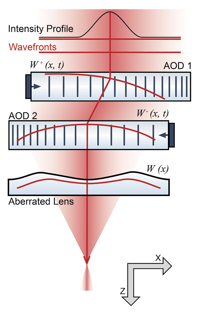
Figure 6. Two acousto-optic detectors (AODs) are combined to act as a single-axis (cylindrical lens) via counter-propagating chirped radio-frequency waveforms. Four can then act as a spherical lens. Courtesy of Angus Silver Lab/UC London.
In the University College London setup, the researchers used commercial AODs provided by G&H and drove them with a high-speed field-programmable gate array-based, home lab-built control system. This generated a range of precise, nonlinear drive frequencies, which were used to shape the optical wavefront. Several years ago, these researchers had already demonstrated continuous line scanning of a laser beam in both the lateral and axial directions using this method, which has the capacity to capture sparsely distributed brain activity. More recently, the researchers have shown that two acousto-optic lenses (with four AODs) can be combined and operated in this way to act as a corrected spherical lens, which could potentially be used in applications beyond fluorescence microscopy, including ophthalmology.
Important applications in neuroscience are pushing the performance envelope of 3D microscopy techniques. Fortunately, the ingenuity of photonics companies and end users is providing innovative solutions that could successfully deliver the requisite performance improvements needed in both speed and imaging depth. In neuroscience, for example, in the time it took to read this sentence, a complex exquisite sequence of electrochemical actions have occurred in the reader’s brain. By imaging similar activities in the mouse brain in real time, fluorescence microscopy could reveal the mysteries how the brain works.
Meet the author
Lars Sandström is vice president of life sciences for the G&H Group, a photonics engineering group that provides differentiating components and subsystems used in a multitude of customer systems. He has more than 30 years of experience in photonics-enabled instrumentation, including lasers and optics; email: lsandstrom@gandh.com.
Acknowledgments
The author would like to acknowledge the following people for their helpful discussions: Paul Kirkby of University College London (UCL) and Agile Diffraction (a UCL spinoff), Çaglar Ataman of Phaseform GmbH, Julien Charton of ALPAO in France, and Gergely Katona of Pázmány Péter Catholic University and Femtonics Ltd.
Reference
1. T. Geiller et al. (2020). Large-scale 3D two-photon imaging of molecularly identified CA1 interneuron dynamics in behaving mice. Neuron, Vol. 108, No. 5, pp. 968-983.e9, www.doi.org/10.1016/j.neuron.2020.09.013.