Dr. Edward Freniere and Michael Gauvin, Lambda Research Corporation
Optomechanical design software helps develop efficient and effective biomedical product designs.
The interdisciplinary nature of optical modeling for life sciences applications implies a high degree of collaboration among medical doctors, life scientists and biomedical design engineers with collective specialties in medicine, optics, mechanics, materials, chemistry and biology. The collaborative efforts of scientists and engineers across these areas require efficient, accurate applications to reduce costs and improve efficacy. Additionally, medical device manufacturers are facing pressure to accelerate time to market while dealing with increasingly tighter R&D budgets and significantly higher cost barriers for certification.
To advance fundamental research and realize product innovation in biomedical optics, software tools can not only facilitate biomedical optical design, but also instill an iterative process allowing engineers to optimize products for the best application-specific results – such as tissue characterization for noninvasive procedures. Optomechanical design software provides a high-productivity environment for developing effective biomedical optical products.
This kind of software tool will lead the designer through a process to maximize the technical merits of the device or system in the shortest amount of time. It minimizes the learning curve, decreases development costs for new products and saves valuable time while offering visual and quantitative feedback.
Modeling light propagation
One such tool, TracePro, is used for modeling the propagation of light in imaging and nonimaging optomechanical systems. Models are produced by creating solid geometry directly within the program or by importing geometry from a lens design or CAD program. Sources can be defined as grids of rays, surface sources or ray files. Rays can propagate through the model nonsequentially with portions of the flux for each ray allocated according to the absorption, specular reflection and transmission, and scattering characteristics of the surface property as defined by the user.
The designer using this tool is not required to be an optical expert to precisely simulate optical systems and produce expedient and accurate models; it includes biomedical properties for tissue and fluorescence applications, enabling both engineers and scientists to work together to produce sophisticated optical models for life sciences applications.
Using such software, a designer can analyze:
• 2-D and 3-D light distributions in illumination and imaging systems
• Stray light, scattered light, and aperture diffraction
• Throughput, loss, or system transmittance
• Flux absorbed by surfaces and bulk media
• Light scattered in biological tissue
• Crosstalk problems in multiple-path systems
• Polarization effects
• Fluorescence effects
• Birefringence effects
Model representation, design work flow
Users can define and manage spectral and angular data for each unique source and combine different sources into a single simulation. To enhance ray-tracing performance, multithreaded algorithms process ray traces in parallel. After ray tracing is complete, a plethora of visualization and analysis capabilities are available to analyze light distribution and fluorescence effects at any point in the optomechanical system. The built-in interactive optimizer is specifically devised for nonsequential design of LED-based biomedical systems and has been proved to reduce design time dramatically compared to traditional trial-and-error prototyping methods. The 2-D and 3-D optimizers feature a sketch utility for quick design entry, interactive ray tracing for design verification, and a downhill simplex optimizer with interactive entry of target specifications.
In addition, a user-configurable system tree creates assemblies, and standard drag-and-drop operations arrange parts and assemblies. Two new irradiance options to enhance imaging are rainbow palette and normalization for identification of stray-light problems in medical devices.
2-D and 3-D optimization
The tool’s 2-D symmetric and 3-D nonsymmetric optimizers are set up with productivity and utility in mind. It allows interactive sketching to quickly enter 2-D and 3-D geometry profiles to create surfaces and then extrude, revolve and combine these surfaces to create sophisticated geometry such as light pipes, biconic reflectors and free-form optics.
Three-dimensional visualization plots allow users to visually track the propagation of light through any ray-traced system with complete pan, zoom and rotational capability using the mouse. The detailed graphics offer new insight and understanding as to how energy and color propagate through an optical system, providing in-depth views to identify unwanted light, stray light and uniformity issues.
Collaborative development
Optical and mechanical assemblies may be designed, analyzed and toleranced using a combination of commercially available lens design software and SolidWorks 3-D mechanical design software. Once imported into the software, lens designs (including parameterized geometry), tolerances, optical materials and CAD geometry are captured in a single model-definition file that can be used and modified. Physical and optical properties are thus encapsulated in one place.
Biological samples, fluorophores, light sources, detectors, coatings and filter data can be specified. The software then performs system-level modeling, analysis and optimization of light distributions, stray light, throughput, flux absorbed by surfaces and bulk media, polarization effects and fluorescence effects.
Libraries of commercially available optical components, light sources, detectors, fluorophores and phosphors, biological tissue and mechanical surface properties are also available or can be customized and added by the user.
TracePro can also be used in complement with another simulation and design program by Lambda Research called OSLO; the two work in tandem for the development of advanced medical systems. OSLO enables a user to design and analyze simple to complex optical models, especially for lenses, mirrors, diffraction gratings, Fresnel lenses and holographic optical elements. The two programs have been successfully used for multiple biomedical applications, including:
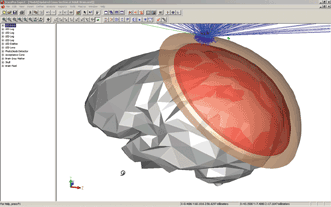
LED emission into the brain to determine oxygenation in tissue. Images courtesy of Lambda Research Corp.
• Developing pulsed light systems like the StarLux and Medilux systems by Palomar Medical Technologies Inc. to reduce varicose veins, to treat acne, wrinkles and vascular lesions, and to remove scars and tattoos.
• Locating and eliminating early cancer in the lungs, colon, cervix and other tissues using fluorescence, laser ablation, photodynamic therapy and other techniques.
• Measuring glucose through optical measurements of skin and blood samples using LED and photodiode sensing systems.
• Developing oximeters to assess blood oxygenation in the brain and other tissue.
• Detecting an object within a tissue (e.g., a tumor) or mapping of functional status within a tissue (e.g., blood perfusion).
Fluorescence
A designer can model fluorescence by importing absorption and emission curves, extinction coefficients and quantum efficiency values from stock fluorophore catalogs or input from proprietary data. Additionally, the designer can enter concentration and wave band of interest. The software calculates excitation efficiency, path length and absorption, and propagates fluorescence-emission rays through the model. It also analyzes light distribution, scatter and fluorescence effects at any point in the optomechanical system.
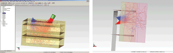
LED emission into human tissue with bulk scatter shown: (left) is the same as (right), except that the setup in (right) has a paddle to hold the LED and detector.
Material properties can be created from actual measured excitation and emission spectra, extinction coefficients and quantum efficiencies, or by applying material properties from the program’s libraries of Invitrogen and Clontech fluorophores.
Light interaction with biological tissue can be simulated using a choice of bulk scatter phase functions; energy propagation can be viewed through tissue with the volume flux viewer. Slice a volume along any axis and analyze it for absorbed, incident, originated or exiting radiation. Designers can use a human tissue catalog or enter unique tissue characteristics, including fluorescence properties.
Design simulation at work
A team of faculty and students at the University of South Florida Colleges of Engineering and Medicine, working with Tampa General Hospital, used TracePro simulations to design state-of-the art laparoscopic endoscopic single-site (LESS) procedures for minimally invasive abdominal surgery. Their innovative research is the first step in developing semiautonomous, wirelessly controlled and networked laparoscopic devices. Their design and implementation of a MARVEL (miniature anchored robotic videoscope for expedited laparoscopy) and camera module will optimize LESS surgery through the self-contained, cable-free MARVEL platform, streamlining the surgical process.
Meet the authors
Dr. Edward Freniere is president and Michael Gauvin is vice president of sales and marketing at Lambda Research Corp.; email: [email protected].
Optical simulation software such as TracePro has enabled innovation and research discoveries in:
• Medical imaging and endoscopy
• Flow cytometry and cell imaging
• Medical imaging
• Pulse oximetry
• In vitro diagnostics
• Biosensors
• Molecular spectroscopy
• Microscopy
• In vivo diagnostics
• Laser and LED surgical devices
Medical product design, evaluation and analytic applications of optical simulation software include:
• Laser- and LED-based surgical devices
• Laser beam delivery systems for surgical instrumentation
• Light distribution devices for both in vivo and in vitro illumination systems
• Reducing stray light and scattered light in biomedical devices
• Studying throughput loss or system transmittance
Analytical applications specific to the life sciences include:
• Tissue propagation
• Glucose monitoring
• Heart rate monitoring
• Fluorescence, Raman, UV, VIS, NIR and IR spectroscopy
• Flow cytometry
• Microarrays and plate readers
• Nucleic acid amplification
• Assay, cell and tissue-based imaging
• Confocal laser scanning and fluorescence microscopy
• Medical imaging and endoscopy
• In vitro and in vivo diagnostics
• Biosensors
• Molecular detection: quantum dots, nanocrystals and luminescent reporters
• Laser-induced fluorescence (LIF) detection
• Förster resonance energy transfer (FRET)
• Flux absorbed by surfaces and bulk media
• Light scattering in biological tissue